Introduction
Cancer remains one of the world’s leading health burdens. Early detection continues to be the strongest determinant of clinical outcomes. Radiologists face rising scan volumes, complex image variations, and growing pressure for rapid reporting. These demands increase the risk of delayed or missed findings, especially in high-throughput environments.
To support this frontline, we introduce the OncoDetect Radiology Accelerator, a next-generation multimodal, AI-augmented imaging companion engineered to elevate abnormality detection such as cancer in chest X-rays (CXRs). The accelerator combines advanced multimodal models, intelligent workflow engines, and clinically aligned automation to reshape how radiologists interact with imaging data. It enhances oncology imaging for AI-powered abnormality detection.
Why Radiology Needs Intelligent Assistance
Medical imaging is entering a new era shaped by foundational AI research. Expanding CXR datasets, vision-language models, and multimodal fusion create new opportunities for precision diagnostics. Within this context, an AI-enabled assistant addresses several persistent challenges:
- Greater Diagnostic Precision: Convolutional Neural Networks (CNNs) trained on extensive datasets deliver state-of-the-art anomaly detection performance, surpassing traditional computer-aided detection methods.
- Relief From High-Volume Workloads: Automating routine tasks lets radiologists focus on ambiguous or high-risk cases, balancing efficiency with accuracy.
- Earlier Identification of Disease Signals: Rapid pattern recognition, supported by large training corpora, increases the likelihood of detecting early-stage oncological indicators when intervention works best.
- Streamlined Clinical Flow: Prioritized case handling and intelligent triage reduce bottlenecks, surface urgent findings sooner and lower turnaround times.
The OncoDetect Radiology Accelerator: How it Works
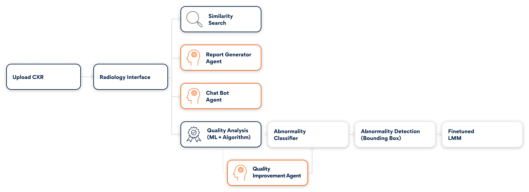
Designed to blend with existing clinical systems, the accelerator strengthens each stage of the diagnostic journey, as seen in Fig 1 and outlined below –
1. Intake & EHR Contextualization
Doctor-patient clinical conversations are transcribed and converted into structured information. The system enriches this context with risk insights from comparable historical records. Radiologists receive a clear patient snapshot before imaging review begins.
2. Feature Embedding of Chest X-Rays
The uploaded CXR is transformed into a high-dimensional embedding through radiology-trained foundation models. These embeddings:
- Capture micro-patterns and texture-level details
- Enable rapid similarity searches across archived cases
- Provide comparative insights that support clinical review
An integrated large language model (LLM) summarizes differential considerations by referencing visually similar examples and offers an evidence-backed perspective.

3. Image Quality Evaluation & Enhancement
A hybrid scoring engine—combining supervised ML and algorithmic rules—evaluates image sharpness, exposure, noise levels, and anatomical coverage. If quality falls short, an enhancement pipeline powered by OpenCV algorithms refines clarity and contrast before analysis continues.
4. Preliminary Abnormality Screening
A foundational embedding model with an MLP head classifies the images based on the likelihood of abnormalities.
This early screening layer helps:
- Prioritize urgent cases
- Reduce manual workload
- Guide radiologists to cases that require deeper review
5. Precise Abnormality Localization
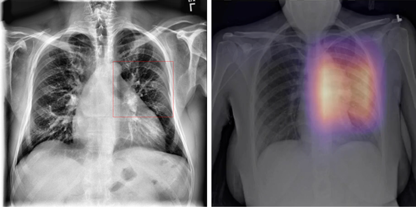
For images flagged as abnormal, a Faster R-CNN model highlights suspicious regions within the lungs. Bounding boxes and heatmaps focus attention on nodules, opacities, masses, and other radiographic abnormalities.
6. Multimodal Explainability & Reasoning
Fine-tuned variants of MedGemma and PaliGemma deliver clinically aligned multimodal explainability. These models produce:
- Justifications for abnormality predictions
- Descriptions of visual cues influencing model confidence
- Interpretable explanations aligned with radiology language
This provides radiologists clear, clinically relevant reasoning behind AI outputs.
7. Interactive Knowledge Assistant
A conversational assistant enables radiologists to query findings, retrieve institutional guidelines, and reference curated medical knowledge bases through Retrieval Augmented Generation (RAG).
Conversations can flow into draft reports, reducing documentation effort.
8. Intelligent Report Generation
Draft impressions generated by LMMs appear in an editable format.
Once validated by the radiologist, the system converts them into standardized structured reports that align with hospital documentation frameworks.
Impact Across the Care Ecosystem
For Radiologists
- Higher Confidence With Comparative Intelligence: Historical case matching provides a quantifiable second opinion based on statistical patterns.
- Reduced Cognitive Load: Streamlined steps and automated data handling minimize repetitive tasks.
- Clearer Multidisciplinary Communication: Concise AI-generated summaries support collaboration across care teams.
For Patients
- Accelerated Diagnostic Timelines: Faster interpretation enables earlier treatment conversations.
- Better Prognoses Through Early Detection: Detecting abnormalities at an early stage improves long-term outcomes.
- Less Emotional Strain: Shorter wait times for results help reduce anxiety during abnormality screening.
Conclusion
The OncoDetect Radiology Accelerator is designed as an extension of clinical expertise, not a replacement. By integrating AI into the radiologist’s workflow, it handles repetitive tasks, surfaces critical insights, and improves diagnostic clarity. This empowers clinicians to deliver faster, more confident, and more equitable abnormality detection.
As healthcare systems move toward AI-driven imaging solutions, the accelerator supports the shift with precision, efficiency, and a commitment to improved patient outcomes. Persistent remains dedicated to collaborating with medical institutions to enhance and deploy this technology responsibly, transparently, and at scale.
Author’s Profile

Shilpa Ramteke
Senior Data Scientist, Innovation Labs AI Research

Jayesh kognole
Lead Software Engineer, Innovation Labs AI Research